Subcutaneous MCT with Transposition Flap Reconstruction
Signalment: 3.5-year-old, FS Potcake
History:
New mass was noted on the dorsal aspect of the left forepaw, present for 1 week.
Physical exam findings:
9.6 mm x 9.6 mm subcutaneous mass on the dorsomedial aspect of the left carpus. No other abnormalities noted.
Diagnostic and clinical staging tests:
Fine-needle aspirate: poorly exfoliative mast cell tumor (MCT)
CBC: no significant abnormalities
Serum biochemistry: no significant abnormalities
Treatment:
Sentinel lymph node (SLN) mapping was performed prior to surgery using 0.5 ml Omnipaque in each of four peritumoral quadrants followed by radiographs at 0, 1, and 3 minutes. This showed simultaneous contrast enhancement of both the ipsilateral superficial cervical (prescapular) and axillary lymph nodes.
A hook needle was inserted into the left axillary lymph node under ultrasound guidance to assist in intraoperative identification of the lymph node.
Intraoperative SLN mapping was performed with 0.5 ml methylene blue in each of four peritumoral quadrants. A standard approach was used to resect the superficial cervical lymph node. An incision was performed adjacent to the hook needle and this incision was continued deeply to identify and excise the axillary lymph node.
The subcutaneous MCT was resected with 10 mm lateral margins and fascia for deep margins. Primary closure was not possible and the defect was reconstructed with a random subdermal plexus transposition flap.
Outcome:
Low grade II MCT with complete histologic (R0) excision, 4.4 mm histologic tumor-free lateral margins and 3.6 mm deep margins
Local flap failure, possibly because of a bandage-related complication. The flap had partial incisional dehiscence and a surgical site infection at postoperative day 11 but was the flap was intact with no necrosis. The flap still appeared 100% viable on postoperative days 12 and 13; but on postoperative day 14, 100% of the flap had become necrotic. This is a very late time period for a flap to fail and it is unusual for the entire flap to fail, so I suspect that the infection and/or the bandage may have contributed to flap failure in this case.
Complete healing by day 43 with second intention.
Tags: #MCT #SLN #SLNmapping #localflap #transpositionflap #reconstruction #secondintentionhealing #complication

Preoperative sentinel lymph node (SLN) mapping showing afferent lymphatic drainage (arrow heads) towards two different but synchronous SLNs, the superficial cervical (or prescapular) and axillary lymph nodes

Preoperative SLN mapping show two synchronous SLNs, which is not very common compared to a single SLN, the ipsilateral superficial cervical and axillary lymph nodes (arrows).

Dr. Gower, one of our board-certified medical oncologists, using an ultrasound intraoperatively to place a hook wire adjacent to the axillary lymph node to aide in identification of the lymph node for excision. (Normal sized axillary lymph nodes are difficult to find!).

Intraoperative SLN mapping was then done with sterile methylene blue injected peritumorally in four-quadrants around the MCT.

The superficial cervical lymph node was excised using a standard approach. Blue discolouration of the lymph node confirms that this is a SLN, corroborating the preoperative SLN mapping findings.

An incision was then performed adjacent to the hook needle and dissection was continued deeply along the hook needle until the axillary lymph node was identified.

Once identified, the axillary lymph node was extirpated using a LigaSure.

Following extirpation of both SLNs, the MCT was resected with 10 mm lateral skin margins and wider subcutaneous tissue margins and one uninvolved fascial layer for deep margins. In these cases, it is more important, at least in my opinion, to get margins in the subcutaneous tissue because this is the anatomical layer from which this tumur is arising, not the skin.
Note the degree of blue discolouration at the surgery site as a result of the intraoperative SLN mapping with methylene blue. This is normal. It is important to mark your lateral margins with a sterile marker pen prior to doing intraoperative SLN mapping so that your surgical approach is not compromised by the blue discolouration in the surgical field.

Because the wound could not be closed primarily, I planned to use a transposition flap from the dorsal aspect of the antebrachium. I recently lectured at the VetEducation Masterclass in Valencia and one of my colleagues and co-lecturers, Dr. Laurent Findji from AURA Veterinary in the UK, showed us this flap. This wound was perfect for reconstruction with this flap. In this photo, you can see me elevating the skin on the dorsal aspect of the antebrachium, demonstrating how much available skin there is this region.

An incision was performed medially and laterally along the elevated skin, with the length of the flap determined by the size of the wound defect.

The flap was then transposed 180 degrees into the defect.

Once transposed into the defect, the flap was sutured with a two layer closure.

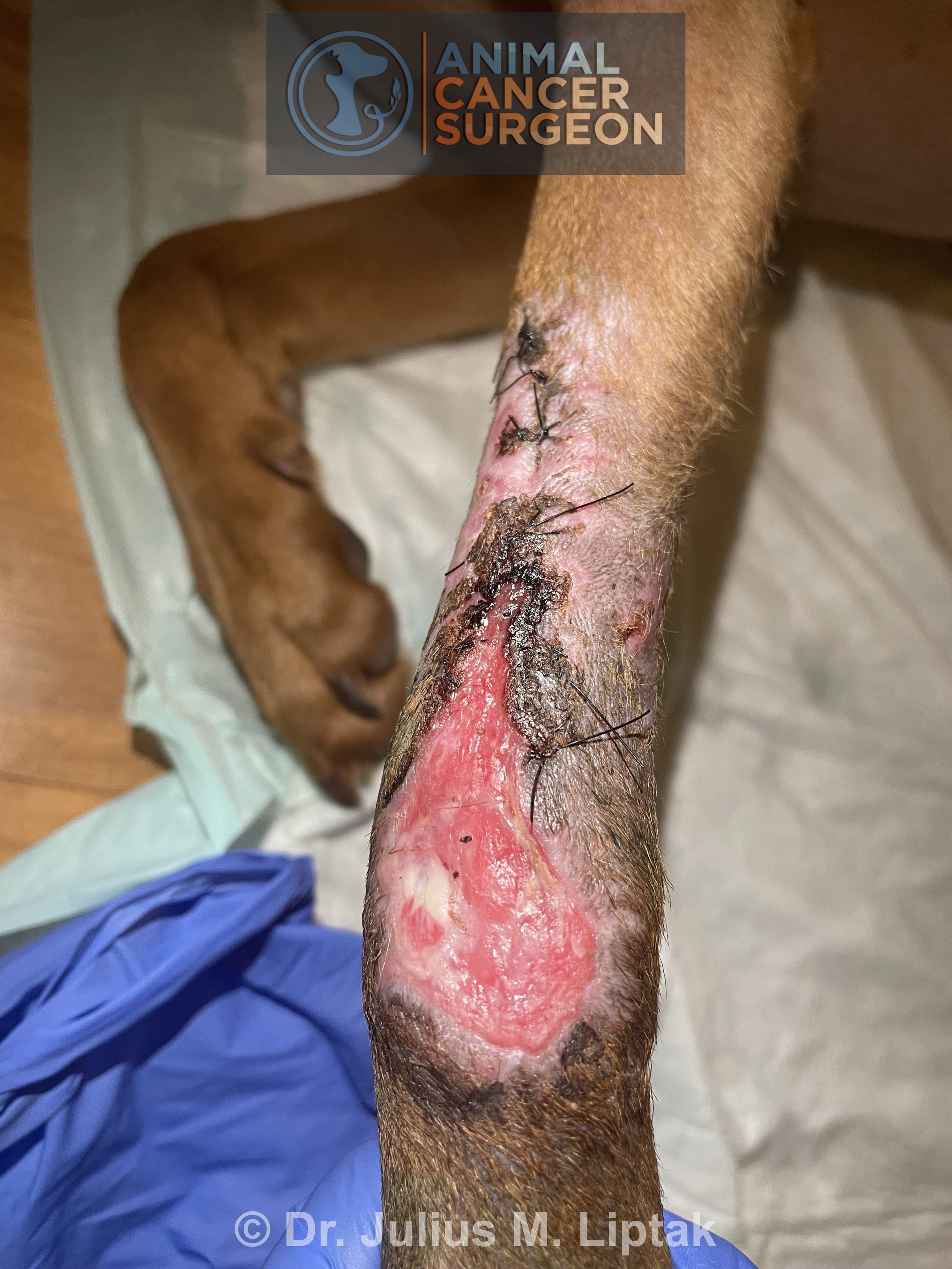
Appearance of the flap on postoperative day 1. The blue discolouration of the flap was residual staining from the methylene blue. The flap remained warm and viable despite this appearance.

The flap developed acute and complete necrosis on postoperative day 14. The wound was initially managed with wet-to-dry bandages. This is the appearance of the wound on day 2 of second-intention healing, with a healthy granulation tissue bed developing.
Day 9 of second-intention healing. The wound was initially managed with daily bandage changes using a non-adherent Telfa pad as the contact layer. The wound has undergone some contraction but minimal epithelialization at this stage.

Day 17 of second-intention healing. The rate of contraction is beginning to accelerate as the wound is visibly smaller.

Day 23 of second-intention healing. The wound is contracting nicely. I forget the actual timing but we have likely changed to bandage changes every 2 days by this time and will soon start using Dermagel, a hydrogel, as the contact layer.

Day 30 of second-intention healing. The wound is contracting beautifully. It is interesting how some wound will initially be slow to start healing but then they go through a rapid phase of healing, such as in this dog.

Day 37 of second-intention healing. Almost there. By this stage, the owner is doing bandage changes every 4 days and continuing with DermaGel as the contact layer. The wound is likely epithelializing under the scabs that have formed over the incision in areas.

Day 43 of second-intention healing. All healed. Yay!
A few points:
This was a subcutaneous MCT (despite being read out using a grading scheme only validated for cutaneous MCTs). Most subcutaneous MCTs have a more benign biological behaviour compared to cutaneous MCTs. In one study of 306 dogs with subcutaneous MCTs, the metastatic rate was only 4% and the local recurrence rate was only 8% (despite 56% of these MCTs being incompletely excised with a 2% and 12% local recurrence rate for completely and incompletely excised MCTs, respectively). The median survival time was not reached in this study with estimated 1-, 2-, and 5-year survival times of 93%, 92%, and 86%, respectively, with surgery alone. Decreased survival times was reported with increased mitotic rate, infiltrative growth pattern, and presence of multinucleated cells. Because of the less aggressive nature of many subcutaneous MCTs, surgical resection can be more conservative with a good chance of success.
The complication rate following reconstruction with a subnormal plexus flap, such as this transposition flap, is 51%.
Second-intention healing can be used as a rescue option for failed reconstructive surgery or can be used as a first-line option to manage an open wound following a tumor resection instead of using a reconstruction option.
Second-intention healing is a labour intensive option. Owners are typically shown how to bandage changes, as was the case with this dog. I encourage owners to email me after each bandage change with a photo of the wound and an assessment of the wound, as well as how they are going with the bandage changes. This allows me to provide information on when the contact layer should be changed and when the frequency of bandage changes can be extended. The owners and I become quite familiar by the end of this process! This dog’s owners did a fantastic job in bandaging and managing her wound, as evidenced by a completely healed wound in less than 6 weeks.