mammary tumors
BACKGROUND
Mammary tumors are the most common tumor in female dogs. Mammary tumors are more common in non-neutered female dogs and cats. The life-time risk of developing a malignant mammary tumor is over 25%. This risk is significantly reduced with early neutering with 0.5% of dogs developing mammary tumors if neutered before their first estrus, 8% between their first and second estrus, and 26% after their second estrus.
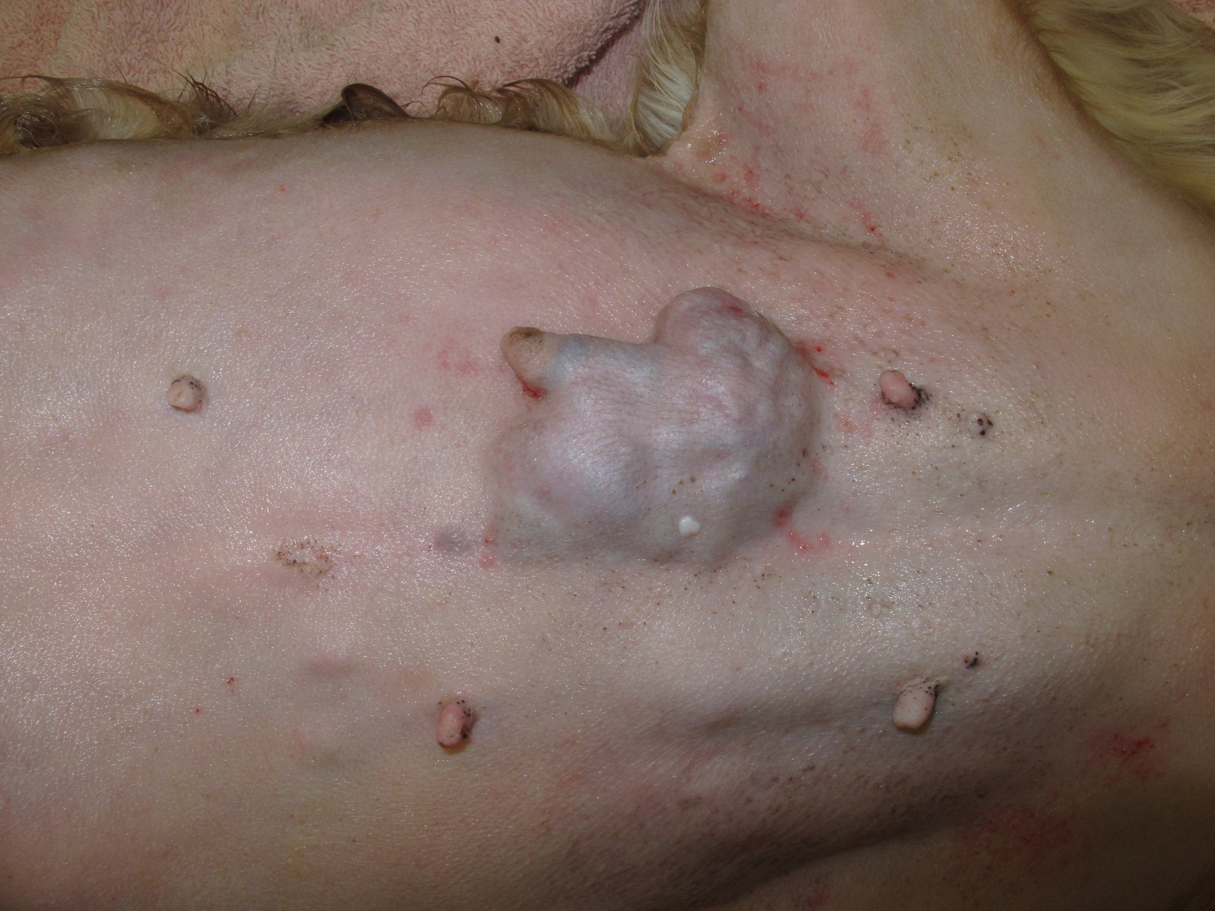
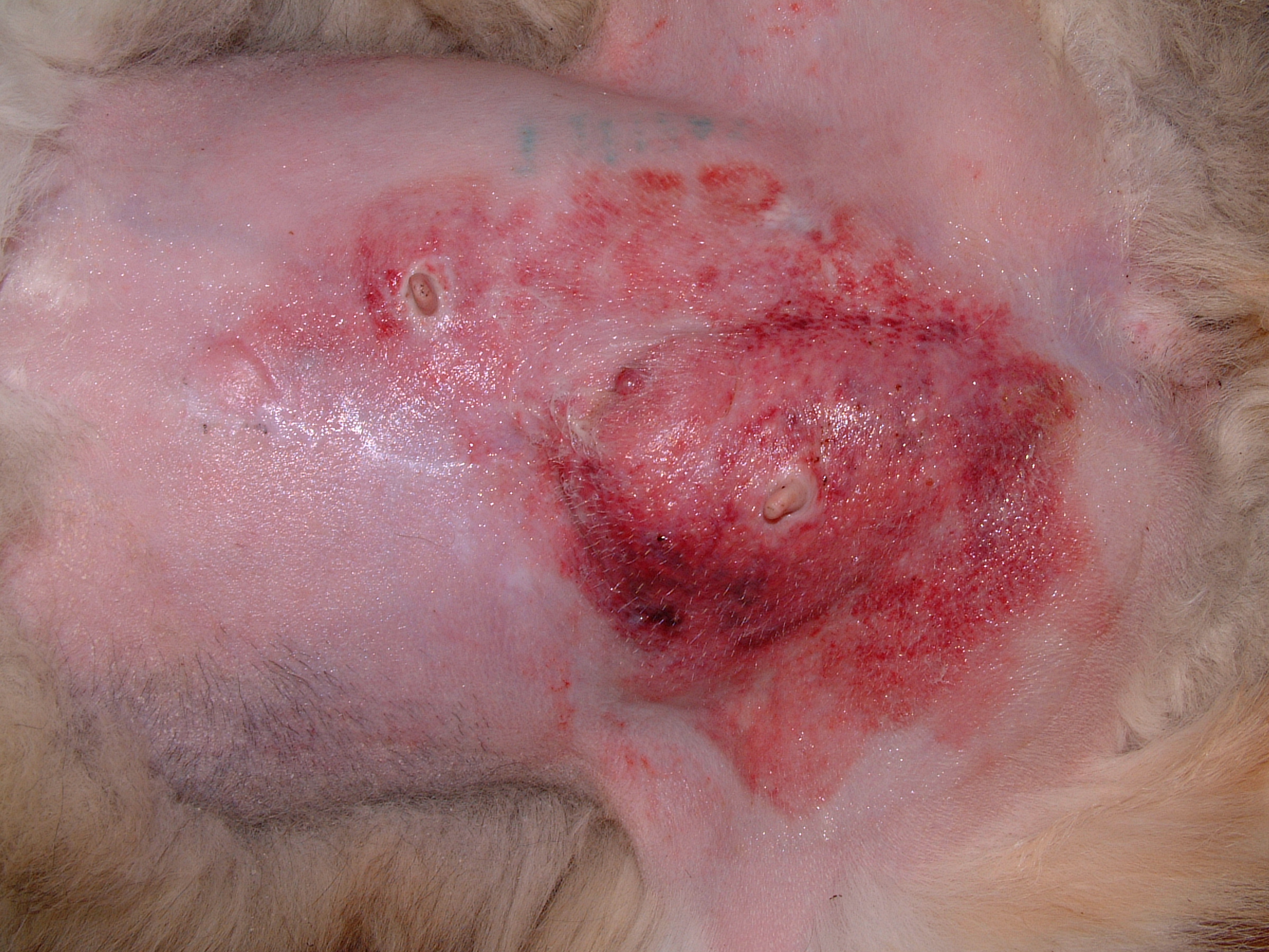
Approximately 50% of mammary tumors in female dogs are benign and 50% are malignant. In contrast, the vast majority of mammary tumors in cats and male dogs are malignant. A variety of malignancies are reported in cats and dogs including carcinomas, inflammatory carcinomas, sarcomas, and carcinosarcomas. Multiple mammary tumors are relatively common in dogs and the fourth and fifth mammary glands are most frequently affected. Benign mammary tumors are often small, well-circumscribed and firm. Malignant mammary tumors are usually rapidly growing with ill-defined borders and frequent inflammation and/or ulceration.
DIAGNOSIS
Mammary tumors are often suspected based on location. Fine-needle aspirates can be performed, but cytology rarely differentiates between benign and malignant mammary tumors and surgical treatment is the same in dogs regardless of whether the tumor is benign or malignant.
CLINICAL STAGING
The two most common sites for metastasis are the regional lymph nodes and lungs. The regional lymph nodes should be palpated and aspirated. Sentinel lymph node mapping should be considered because the sentinel lymph node (which is defined as the first draining lymph node) may be different to the regional lymph node. Sentinel lymph node mapping involves peritumoral injection of a lipid-soluble contrast agent 24 hours prior to surgery (usually under sedation), regional radiographs immediately prior to surgery to identify the sentinel lymph node, and then peritumoral blue dye injection during surgery to aide in intraoperative identification and excision of the sentinel lymph node. Alternatively, if unilateral or bilateral mastectomy is planned, then just intraoperative peritumoral blue dye is often sufficient for the identification of the sentinel lymph node. Thoracic radiographs or CT scans are recommended to assess the lungs for metastasis.
TREATMENT
Dogs
Surgical excision is recommended for treatment of mammary tumors in both cats and dogs. In dogs, the aggressiveness of surgery does not determine prognosis and depends on the size, location and number of tumors. Most tumors can be excised with 1 cm margins. Alternatively, unilateral mastectomy can be considered as a therapeutic and preventine procedure as up to 60% of dogs will develop further mammary tumors in the ipsilateral mammary chain following local mastectomy. Neutering at the time of mastectomy is recommended in both cats and dogs, although the effect of this on prognosis is controversial. Neutering likely has the greatest effect on benign and low-grade malignancies as these tumors will often have either estrogen and/or progesterone receptors, whereas these receptors are absent in more aggressive malignant mammary tumors.
The need for chemotherapy is determined by the tumor type, but remains controversial in dogs with mammary carcinomas. Only two studies have shown a survival benefit with the use of chemotherapy postoperatively, and one study included only eight dogs and in the other study chemotherapy was only effective for dogs with stage IV disease (lymph node metastasis). Larger and more recent studies have shown no benefit on survival time with the addition of chemotherapy, regardless of clinical stage, histologic grade, or the presence of lymphatic invasion. While a survival benefit has not been shown conclusively, chemotherapy should be considered for high-grade malignant mammary tumors, mammary tumors with evidence of vascular or lymphatic invasion, and mammary sarcomas because of the high risk of metastatic disease. In one clinical paper (2016), while not significant because of low case numbers, the combination of carboplatin and mitoxantrone in an alternating protocol seemed to hold some promise.
Cats
In cats, the prognosis is significantly improved with a more aggressive bilateral mastectomy (or excision of both mammary chains). Bilateral mastectomy can be performed in a single stage or can be staged by 4-6 weeks. There is a significantly increased risk of complications following single-stage bilateral mastectomy in cats, but the majority of these complications are minor wound complications which can be managed medically or with minor revisions.
Postoperative chemotherapy is recommended in cats as a soon-to-be published paper of 126 cats with mammary carcinomas treated surgically showed that chemotherapy significantly improves survival time in these cats.
PROGNOSIS
Dogs
The prognosis in dogs is dependent on clinical presentation, clinical stage, surgical margins, and tumor type. Benign mammary tumors are cured with surgical excision (although the development of new and unrelated tumors is reported in up to 60% of dogs). Clinical staging is used to describe how advanced malignant mammary tumors are with stage I being a non-metastatic mammary tumor < 3 cm in diameter; stage II is a non-metastatic mammary tumor 3-5 cm in diameter; stage III is a non-metastatic mammary tumor > 5 cm in diameter; and stage IV and V are mammary tumors with lymph node and distant metastasis, respectively, regardless of size of the mammary tumor. The following are the median survival times and 1- and 2-year survival rates for each clinical stage of mammary carcinoma in dogs:
- Stage I: median survival time 500-1226 days, and 1- and 2-year survival rates of 61.3% and 61.3%
- Stage II: median survival time 420-924 days, and 1- and 2-year survival rates of 60.0% and 60.0%
- Stage III: median survival time 210-287 days, and 1- and 2-year survival rates of 30.8% and 10.3%
- Stage IV: median survival time 90-165 days, and 1- and 2-year survival rates of 28.8% and 14.4%
- Stage V: median survival time 224 days, and 1- and 2-year survival rates of 43.8% and 29.2%
Prognostic factors associated with clinical presentation include the number of tumors, presence of ulceration, and fixation of tumors to underlying tissue. Survival outcome is significantly worse for dogs with ulcerated mammary carcinomas with a median survival time of 118 days and 1- and 2-year survival rates of 10.8% and 5.4%, respectively, compared to a median survival time of 443 days and 1- and 2-year survival rates of 52.6% and 45.0%, respectively, for dogs with non-ulcerated mammary carcinomas.
The use of desmopressin in the perioperative period significantly improves median survival times in dogs with grade II and III mammary carcinomas. In one prospective study of 18 dogs treated with desmopressin and 10 dogs treated with a saline placebo, the overall median survival time was significantly longer in dogs treated with desmopressin (809 days) compared to dogs in the placebo group (237 days) with 17% and 80% of dogs dying because of disease-related reasons in each group, respectively. Furthermore, the median survival times for dogs with grade II and III mammary carcinomas in the placebo group were 351 days and 35 days, respectively, compared to > 600 days when treated with desmopressin.
In a recent clinical study (2016) of 94 dogs with mammary carcinomas, the completeness of excision was the strongest predictor of survival. The median survival time for dogs with incompletely excised mammary carcinomas was only 70 days with 1- and 2- year survival rates of 14.8% and 0%, respectively. In comparison, median survival time for dogs with completely excised mammary carcinomas was 872 days with 1- and 2- year survival rates of 56.2% and 51.3%, respectively. The effect on histologic margins was maintained for dogs with stage I-III mammary carcinoma (but not stage IV and V mammary carcinomas) and mammary carcinomas with lymphatic invasion. The median survival time for dogs with incompletely excised stage I-III mammary carcinomas was only 68 days with 1- and 2- year survival rates of 12.5% and 0%, respectively; compared to a median survival time of 1098 days with a 66.1% 1- and 2- year survival rate. The median survival time for dogs with mammary carcinomas with histologic evidence of lymphatic invasion and incomplete excision was only 70 days with 1- and 2- year survival rates of 16.7% and 0%, respectively; compared to a median survival time of 347 days with 1- and 2- year survival rates of 44.5% and 29.7%, respectively, for dogs with completely excised mammry carcinomas with lymphatic invasion.
Histologically, the presence of lymphatic invasion and vascular invasion and potentially histologic grade have prognostic significance for dogs with mammary carcinomas. In a recent clinical study (2016), the median survival time for dogs with histologic evidence of lymphatic invasion was 179 days with 1- and 2-year survival rates of 29.1% and 13.2%, respectively. In comparison, the median survival time for dogs with no evidence of lymphatic invasion was 1098 days with 1- and 2-year survival rates of 65.7% and 62.9%, respectively.
Tumor-related mortality varies from 15% for adenocarcinoma, 65% for ductular adenocarcinoma, and 100% for inflammatory carcinoma, carcinosarcoma, and sarcoma. The median survival times for various tumor types are 25-60 days for inflammatory carcinomas, 90 days for mammary osteosarcomas, 6 months for other mammary sarcomas, and 2.5 months for anaplastic carcinomas, 16 months for solid carcinomas, and 21 months for adenocarcinomas.
Cats
The prognosis in cats is dependent on clinical stage, surgical aggressiveness, and degree of differentiation. Stage I is a non-metastatic mammary tumor < 2 cm in diameter; stage II is a non-metastatic mammary tumor 2-3 cm in diameter; stage IIIa is a non-metastatic mammary tumor > 3 cm in diameter; and stage IIIb and IV are mammary tumors with lymph node and distant metastasis, respectively, regardless of size of the mammary tumor. The median survival times for cats with stage I, II, and IIIa mammary carcinomas are 450 to > 1095 days, 448-730 days, and 180-200 days, respectively. The median survival time for cats with stage IIIb disease ranges from 6 months to 1543 days; while the median survival times for cats with lung metastasis and pleural effusion are 331 days and 188 days, respectively. The 1-year survival rates for well, moderately, and poorly differentiated mammary carcinomas are 100%, 42%, and 0%. The median survival times for cats with histologic evidence of local invasion and vascular or lymphatic invasion are 21.8 months and 13.4 months, respectively. The median survival time for cats treated with subtotal mastectomy is 216 days compared to 566 days for unilateral mastectomy and 917 days for bilateral mastectomy. In a recent study of 126 cats, treatment with postoperative chemotherapy significantly improved survival times when combined with bilateral mastectomy.
Last updated on 6th March 2017


