REVERSE SAPHENOUS CONDUIT AXIAL PATTERN FLAP
Axial pattern flaps are pedicle grafts which incorporate a direct cutaneous artery and vein at their base. While not a true axial pattern flap, branches of the saphenous artery are the direct cutaneous artery for the reverse saphenous conduit axial pattern flap. The direct cutaneous artery and vein extend along the length of the flap for a variable distance and the terminal branches supply the subdermal, cutaneous, and subpapillary plexuses. The reverse saphenous conduit axial pattern flap is indicated for reconstruction of wounds of the distal pelvic limb.
In a 2015 study investigating the outcome of axial pattern flaps in 49 dogs and 24 cats, postoperative complications were reported in 89% of animals. The most common complications included dehiscence (50% of axial pattern flaps in dogs and 75% of axial pattern flaps in cats), flap swelling (43% of axial pattern flaps in dogs and 50% of axial pattern flaps in cats), necrosis (46% of axial pattern flaps in dogs and 15% of axial pattern flaps in cats), infection (27% of axial pattern flaps in dogs and 40% of axial pattern flaps in cats), non-infectious discharge (14% of axial pattern flaps in dogs and 45% of axial pattern flaps in cats), and seroma (23% of axial pattern flaps in dogs and 20% of axial pattern flaps in cats). While the complication rate associated with axial pattern flaps is high, all of these complications were managed with either simple revisions (e.g., debridement and resuture) or conservatively (e.g., antibiotics, bandages, or monitoring). Overall, 93% of wounds were successfully reconstructed with an axial pattern flap. The flap outcome was assessed as excellent in 23%, good in 41%, fair in 30%, and poor in 7% of animals. The outcome following reconstruction using a reverse saphenous conduit axial pattern flap was not specifically stated in this study. In a 1983 study, 100% survival of the reverse saphenous conduit flap was reported in 15 dogs. In a 2014 study, the reverse saphenous conduit axial pattern flap was used to reconstruct tarsal defects resulting from either trauma or resection of cancers with a low incidence of postoperative complications (described as necrosis of < 15% of the distal aspect of the flap).
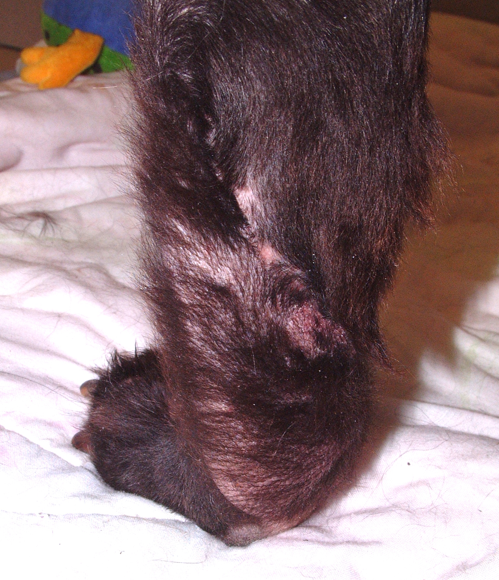
Case 1 - Skin Graft Failure Following Resection of a Cutaneous Metatarsal Soft Tissue Sarcoma
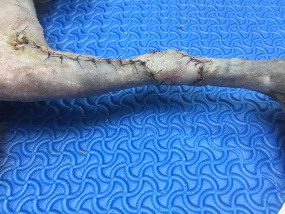
Case 2 - Traumatic Hock Wound (Courtesy Dr. Nithida Boonwittaya)
Last updated on 6th March 2017


