SURGICAL MARGINS
Surgical oncology involves the surgical management of benign tumors and malignant cancers. The treatment of animals with cancer is well-established, and continues to be a growing field as improvements in preventive medicine and diets have resulted in our pets living longer and longer. Cancer is now the leading cause of death in older cats and dogs, and hence there is a need for everyone, from owners to family veterinarians to specialists, to be aware of the options available for the diagnosis and treatment of animals with benign tumors and malignant cancers. There are some important considerations when dealing with masses in animals, including preoperative biopsy, surgical margins, and histopathologic features of the tumor (such as tumor type, histologic grade, and histologic margins).
SURGICAL MARGINS
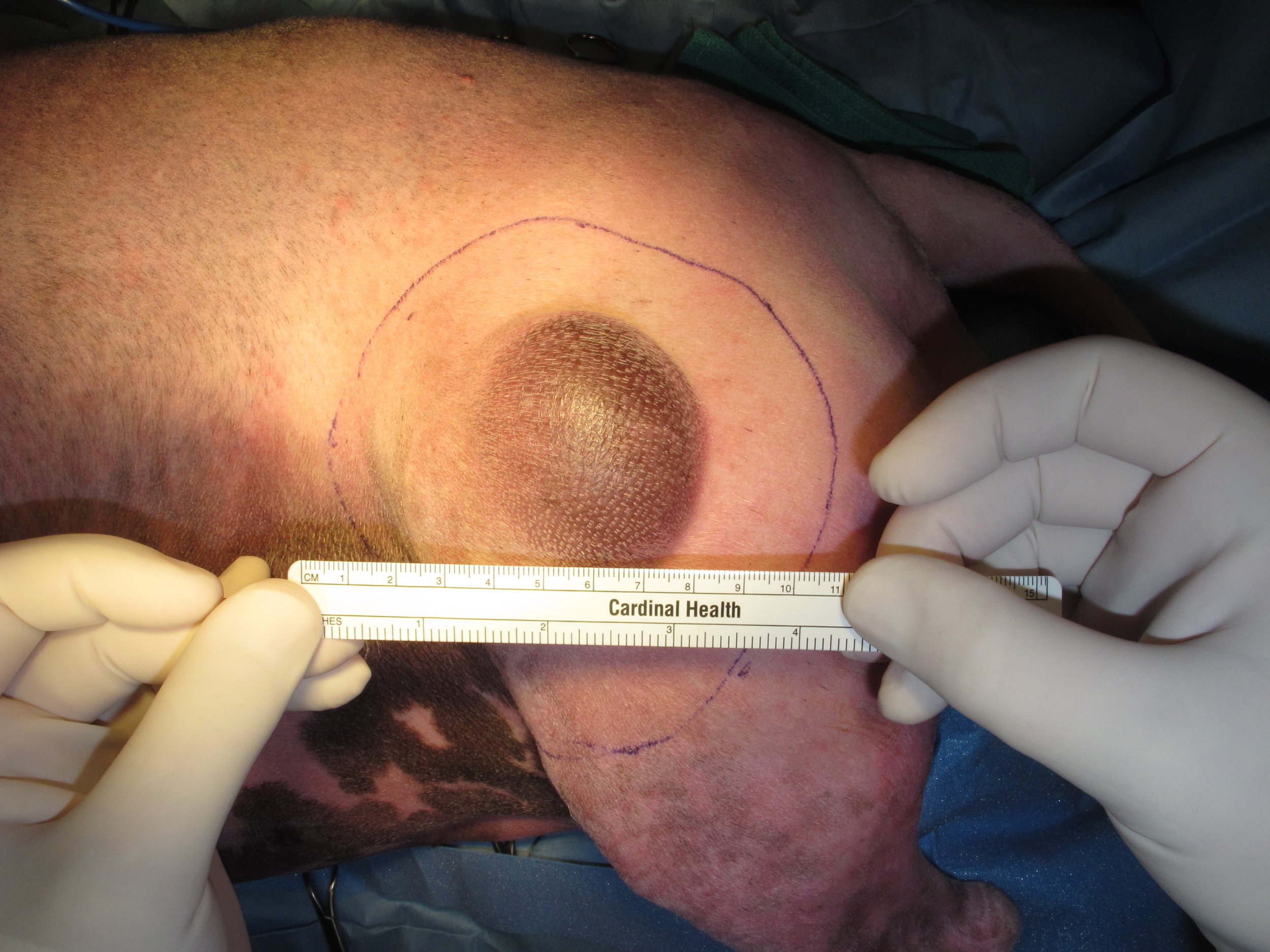
Surgical margins refers to the amount of normal tissue excised around the tumor. Surgical margins are usually defined in terms of metric measurements (lateral margins) and anatomic barriers such as a fascial layer (deep margins). Recommendations for the width of the lateral margins is determined by tumor type: 1cm for carcinomas, 2cm for mast cell tumors, 3cm for soft tissue and bone sarcomas, and up to 5cm for feline injection-site sarcomas. Fascial barriers are used for deep margins as fascia is considered resistant to invasion by cancer cells, unlike other tissues such as subcutaneous fat. These margin recommendations are largely arbitrary and based on personal opinions and experiences. The only tumor for which surgical margins have been studied is canine cutaneous mast cell tumors where 1cm lateral margins result in complete histologic excision of 100% of grade I mast cell tumors and 75% of grade II mast cell tumors, and 2cm lateral margins result in complete histologic excision of 100% of both grade I and II mast cell tumors.
The surgical margin classification scheme used in veterinary surgical oncology has been adapted from the classification scheme proposed by Enneking for musculoskeletal sarcomas in people. The premise of this classiciation scheme is that the tumor is surrounded by a pseudocapsule (reactive zone) which may contain microscopic tumor extensions or satellite populations of tumor cells. While this classification scheme is widely used in veterinary surgical oncology, it has not been used for tumors other than musculoskeletal sarcomas in people and has not been validated for any tumor type, musculoskeletal sarcoma or otherwise, in animals. This classification scheme divides the aggressiveness of surgery, or surgical dose, into four categories: intralesional, marginal, wide, and radical.
Intralesional excision is when the surgical incision is through the tumor and results in gross residual tumor. This approach is rarely, if ever, indicated, even for palliative resections.
Marginal excision is when the surgical incision is through the reactive zone between the tumor and the pseudocapsule. By definition, lateral margins < 1cm for carcinomas, < 2cm for mast cell tumors, < 3cm for soft tissue and bone sarcomas, and 5cm for feline injection-site sarcomas.
Wide excision is when the surgical incision is outside of the reactive zone and pseudocapsule and in grossly normal tissue in all dimensions. By definition, lateral margins 1cm or greater for carcinomas, 2cm or greater for mast cell tumors, 3cm or greater for soft tissue and bone sarcomas, and 5cm or greater for feline injection-site sarcomas.
Radical excision is when a tumor is excised en bloc with the anatomic compartment containing the tumor. For example, for a sarcoma within the quadriceps muscle, radical excision includes resection of the entire quadriceps muscle. This classification has been modified in veterinary surgical oncology with radical excision often referring to wide excision with en bloc resection of a body part (i.e., limb or spleen).
In people, wide and radical excisions are usually adequate for local tumor control (i.e., these approaches are associated with a low rate of local tumor recurrence). The same is true in cats and dogs using the recommended lateral and deep margins described above. In people, the wide excision classification has been further stratified as wide adequate (2cm-5cm lateral margins) and wide inadequate (1cm-2cm lateral margins). In animals, however, the controversies include what margins are defined as marginal or wide in the absence of evidence-based medicine. For example, the dogma has always been that soft tissue sarcomas should be excised with 3cm lateral margins; however there is increasing evidence that lesser margins are adequate for excision of soft tissue sarcomas on the extremities with similar rates of local tumor control. So, in this blurred line between marginal and wide excisions, should a marginal excision be defined as lateral margins < 2mm, 1cm, or 2cm?
Last updated on 6th March 2017


