CAUDAL and hemiMAXILLECTOMY
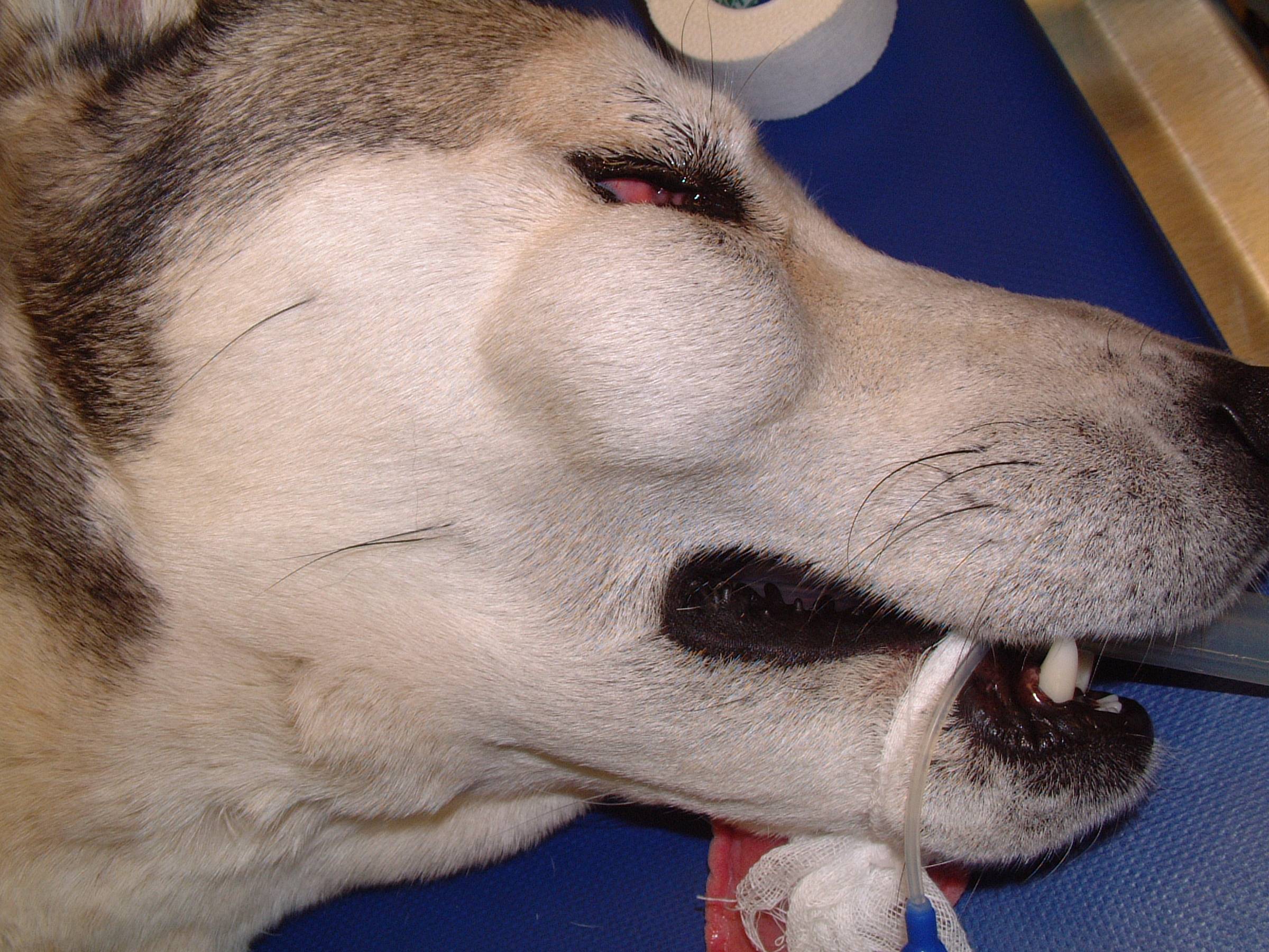
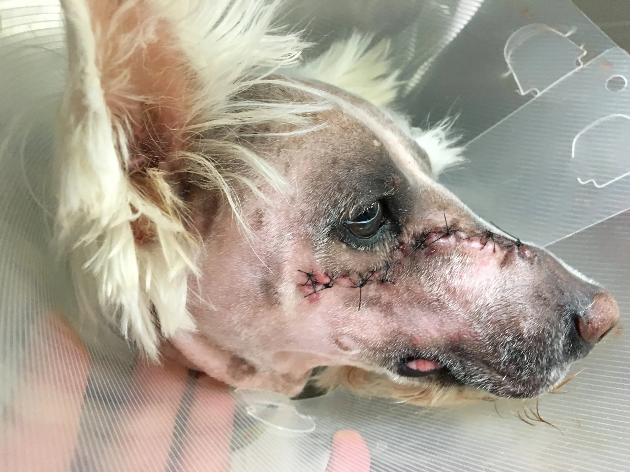
Hemimaxillectomies and caudal maxillectomies are surgical procedures which involve removal of the entire hemimaxilla or the caudal portion of the maxilla, and these can be combined with more extensive procedures (such as inferior orbitectomy, orbitectomy and enucleation, and craniectomy) if required. These surgeries are indicated for extensive or caudally located malignant tumors which have not crossed the midline of the hard palate. There are two approaches to perform hemimaxillectomy and caudal maxillectomy: intraoral and combined dorsolateral skin-intraoral. The intraoral approach is used for tumors confined to the dental arcade, and the combined approach is preferred for tumors which have extended dorsally or caudally from the dental arcade.
The most common complications encountered with hemimaxillectomy and caudal maxillectomyinclude intraoperative bleeding, epistaxis, incisional swelling, and lip ulceration. Intraoperative bleeding is more common with hemimaxillectomy and caudal maxillectomy, either through an intraoral or combined dorsolateral-intraoral approach, than other types of maxillectomies because of transection of the maxillary artery as it courses through the ventral aspect of the inferior orbit. In two older clinical studies (1986 and 1992), intraoperative hemorrhage resulted in the death of one dog (1.4%, 1/69); a blood transfusion was required for the correction of intraoperative blood loss and subsequent hypovolemic shock in a further 12% of dogs (2/17); and a further 9% of dogs (6/69) had blood loss resulting in hypovolemic shock but did not require a blood transfusion. In a more recent clinical study (2003) of caudal maxillectomy via a combined dorsolateral-intraoral approach in 20 dogs, significant blood loss was reported in 75% of dogs with a mean decrease in packed cell volume of 15.4%; and a whole blood transfusion was required in 30% of dogs. Dehiscence of the intraoral wound results in an oronasal fistula and is reported in 5% of dogs treated with either hemimaxillectomy or caudal maxillectomy. Dehiscence is more common in dogs treated with either pre- or post-operative radiation therapy. For dogs with incisional dehiscence, the majority require surgical revision (70%) although conservative management can also be used for smaller dehiscences and those without oronasal fistulae. The lip is drawn towards the midline and the extent of this medialization of the lip will depend on the extent of resection of the hard palate, the length of the lip, and the degree of undermining of the mucosal-submucosal labial flap. Lip trauma and ulceration secondary to interference from the ipsilateral mandibular canine tooth following medialization of the lip has been reported in 10% of dogs undergoing caudal maxillectomy via a combined dorsolateral-intraoral approach. Functionally, dogs and cats do well following hemimaxillectomy and caudal maxillectomy with the vast majority of animals returning to voluntary eating within 1-3 days of surgery. In surveys of owner satisfaction, 85% of owners were satisfied with the functional and cosmetic outcomes following all types of maxillectomies, and 100% of owners were satisfied following caudal maxillectomy via a combined dorsolateral-intraoral approach.
PRE- AND POST-OPERATIVE APPEARANCE - INTRAORAL APPROACH
PRE- AND POST-OPERATIVE APPEARANCE - COMBINED DORSOLATERAL-INTRAORAL APPROACH
Complications
Paradoxical Skin Movement
Paradoxical Skin Movement
Last updated on 6th March 2017


