Incisivectomy in a Dog with an Acanthomatous Ameloblastoma
Signalment: 7-year-old, FS Australian shepherd criss
History:
This dog presented to her family veterinarian with a rapidly growing rostral maxillary mass. Blood work was done and a biopsy was consistent with an acanthomatous ameloblastoma (AA).
Physical exam findings:
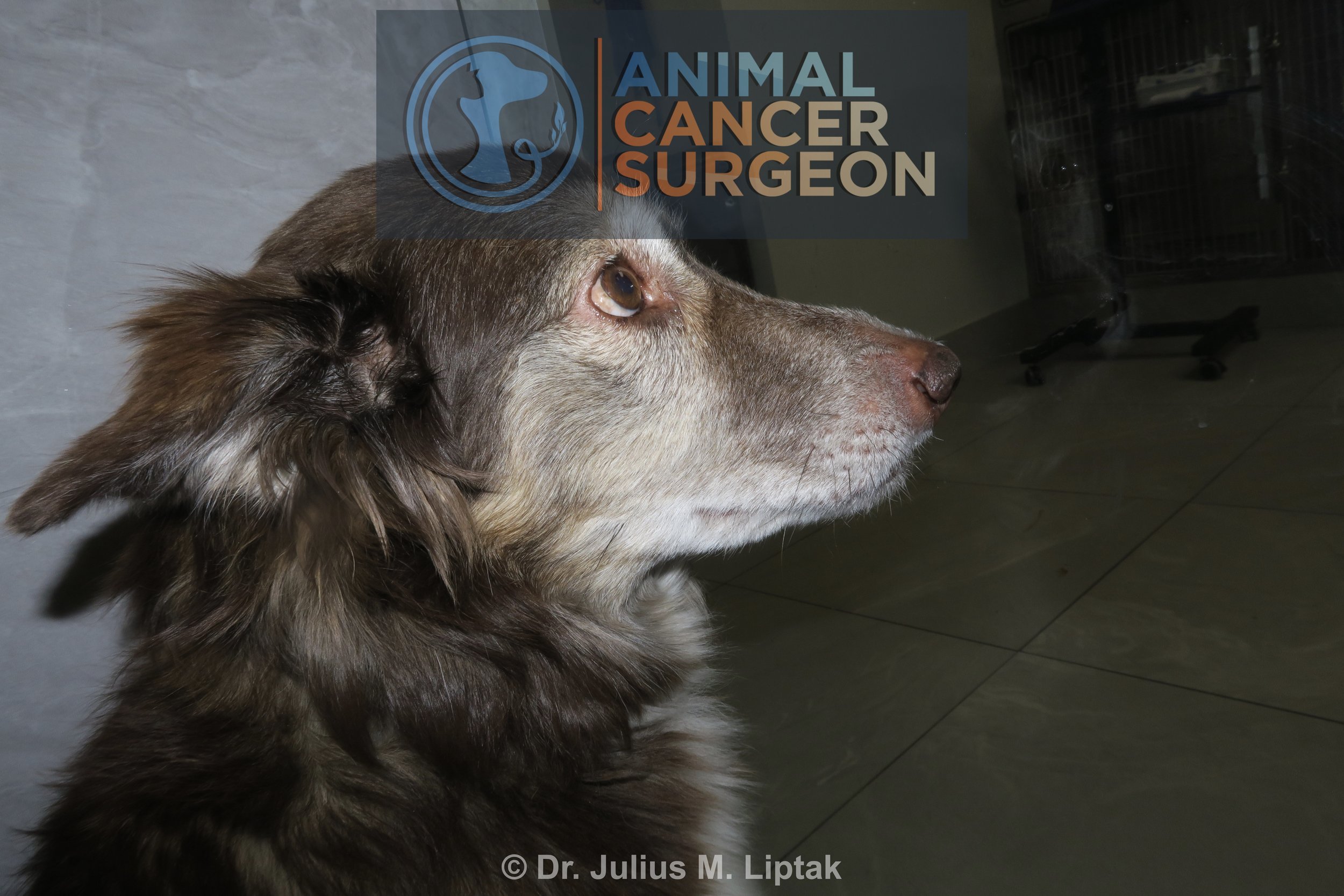
The only abnormality was a raised mucosal covered mass extending from 102 to 203.
Diagnostic and clinical staging tests:
CBC: no abnormalities
Serum biochemistry: increased SDMA with normal BUN and creatinine
Urinalysis: no abnormalities
Biopsy: acanthomatous ameloblastoma
Treatment:
The three treatment options for dogs with AA are surgery, radiation therapy, and intralesional bleomycin. Of these, surgery is usually recommended because it is typically quicker and cheaper and more likely to achieve tumor control. Because of the location of the AA in this dog, the AA was able to be resected with 1 cm caudal margins using a biradial (TPLO) saw to preserve the canine teeth. This procedure is called an incisivectomy.
Outcome:
Acanthomatous ameloblastoma excised with complete histologic margins with the narrowest lateral margin measuring 2.0 mm and the narrowest caudal histologic tumor free margin measuring 4.4 mm.
Minor wound dehiscence which healed by second intention.
This surgery should be curative for this dog as AAs are benign tumors and local recurrence is rare, especially following complete histologic excision.
Video link: https://www.youtube.com/watch?v=1L8E27cYyIc&t=4s
Tags: #CAA #acanthomatousameloblastoma #incisivectomy #maxillectomy

Immediate preoperative image of the acanthomatous ameloblastoma, a benign but invasive oral tumor, arising from the upper incisor region in a dog.

Immediate preoperative image of the acanthomatous ameloblastoma, a benign but invasive oral tumor, arising from the upper incisor region in a dog.

The gingiva mucosa is incised with a number 10 or 15 scalpel blade and then reflected caudally to expose the dorsal aspect of the planned osteotomy.

A 27 mm biradial saw was then pressed against the palatine mucoperiosteum to create an imprint in the mucoperiosteum, and an incision was made through the mucoperiosteum along this imprint. The mucoperiosteum was then reflected caudally with a periosteal elevator, and the incisivectomy was then performed with a 27 mm biradial (TPLO) saw. This is what these saw blades were really designed for!

Following the incisivectomy, the transected roots of the incisor teeth were extracted.

Intraoperative image showing all of the retained tooth roots have been removed.

The mucosal incision was closed in a single layer of 3-0 PDS suture material in a cruciate suture pattern.

Postoperative specimen image with caudal margins inked. This was histologically confirmed as a completely excised (4.4 mm caudal margins), benign acanthomatous ameloblastoma.
Postoperative appearance 1 day after surgery. There is a very mild droop of the rostral nose.